


Les petits détails font une GRANDE différence : Reconnaître et gérer les comportements perturbateurs en milieu Clinique
Les petits détails font une GRANDE différence est un programme d’autoévaluation agréé (section 3) disponible sur ligne.
Cette activité d’apprentissage a été conçue pour aider les médecins en exercice, les résidents, les administrateurs et les autres membres des équipes soignantes à acquérir les connaissances et les compétences nécessaires et à adopter les comportements qui s’imposent pour mieux cerner et régler les problèmes liés au comportement perturbateur de certains de leurs coéquipiers.
La premisse du programme
Cette activité suivre le cas de Mme Wendy Lee, une patiente qui a subi une intervention chirurgicale au Réseau de santé Pondview.
L’intervention chirurgicale de Mme Lee ne s’est pas déroulée comme prévu, et la Dre Gin Staples, l’une des administratrices de l’hôpital, doit examiner son cas en vue de formuler des recommandations. Votre objectif est d’analyser le cas de Mme Lee selon la perspective des membres de l’équipe soignante, afin d’aider la Dre Staples à formuler des recommandations visant à favoriser la création d’une « culture juste » au sein du Réseau de santé Pondview.
Vous devrez répondre à des questions au fur et à mesure que vous prendrez connaissance de ce cas. Veuillez prendre vos réponses en note au fil de votre lecture. Vous trouverez au bas de cette page un corrigé qui vous aidera à évaluer dans quelle mesure vous avez compris la matière. En répondant à ces questions, vous recueillerez des éléments qui serviront de base à la formulation des recommandations finales de la Dre Staples. Vous pourrez également les utiliser dans le cadre d’un projet de formation personnel afin d’obtenir les crédits liés à la section 2 du programme de Maintien de la certification (MDC) auxquels vous avez droit.
Objectifs d’apprentissage
À la fin de cette séance d’apprentissage par simulation, les participants pourront :
- Reconnaître les stimuli environnementaux à l’origine des comportements perturbateurs
- Définir des stratégies pour enrayer les comportements perturbateurs en milieu clinique
- Décrire l’incidence des comportements perturbateurs sur le milieu clinique
- Mettre en pratique des techniques qui favorisent une « culture juste » dans le milieu clinique
- Réfléchir à des cas de comportements perturbateurs en milieu clinique
Crédits de DPC
Il s’agit d’un programme d’autoévaluation agréé (section 3), tel que défini dans le programme de Maintien du certificat du Collège royal des médecins et chirurgiens du Canada, et approuvé par l’Unité du développement professionnel continu du Collège royal. Vous pouvez réclamer un maximum de deux heures (les crédits sont calculés automatiquement).
Détails d’accès
Pour accéder au module, veuillez visiter le site Web du Collège royal des médecins et chirurgiens du Canada.
CanMEDS Program: Better Practice, Better Outcomes
The CanMEDS Framework was developed by the Royal College of Physicians and Surgeons of Canada to provide key strategies for healthcare professionals to increase efficiencies in their practice, improve communication with patients and colleagues, and demonstrate stronger leadership.
The program explicitly expresses how CanMEDS roles are used in everyday clinical and non-clinical practice. The CanMEDS roles have been developed based on what patients believe are the most important competencies and abilities that physicians should possess.
Target Audience
This self-assessment program was developed with local faculty and subject matter experts to ensure a high degree of scientific integrity, rigor and balance for practitioners.
Learning Objectives
After successful completion of the program, participants will be able to:
- Use the CanMEDS Framework to improve your practice and your patient outcomes
- Identify areas for your continuing professional development using the CanMEDS Framework in all seven roles: Communicator, Collaborator, Leader, Health Advocate, Scholar, Professional and Medical Expert
CPD Credits
This program was co-developed by the Royal College of Physicians and Surgeons of Canada and mdBriefCase Group.
This activity is an Accredited Self-Assessment Program (Section 3) as defined by the Maintenance of Certification Program of the Royal College of Physicians and Surgeons of Canada, and approved by the Royal College CPD Unit. You may claim a maximum of 1 hour (credits are automatically calculated).
Access Details
The online learning activity is free for RCPSC Fellows. To access the CanMEDS online self-assessment program, please visit the mdBriefCase website for more information.
Survey of reported eye injuries from handheld laser devices in Canada
Read the full article through the Canadian Journal of Ophthalmology website: Full Article
Authors
Sami S. Qutob, Katya P. Feder, Michelle O’Brien, Leonora Marro, James P. McNamee, David S. Michaud
Abstract
Background
Unprotected exposure to handheld lasers can cause temporary or permanent vision loss depending on the laser classification.
Objective
To evaluate the occurrence of, and details associated with, reported eye injuries resulting from handheld lasers.
Methods
A 14-item questionnaire developed by Health Canada was distributed by the Canadian Ophthalmological Society and the Canadian Association of Optometrists to their respective members.
Results
Questionnaire data were available from 909 respondents (263 ophthalmologists; 646 optometrists). Response rates were 23.1% and 12.7%, respectively. Validated data were available from 903 respondents, where 157 (17.4%) reported encountering at least 1 eye injury from a handheld laser. A total of 318 eye injuries were reported with an annual increase of 34.4% (95% CI 21.6%–48.7%, p < 0.0001) between 2013 and 2017. When respondents reported on only their most severe case, 77 (53.5%) reported vision loss that ranged from minor to severe, which persisted for more than 6 months in 42.9% of the cases. Another 59 (41.3%) noted the presence of retinal damage. The prevalence of eye injuries from handheld lasers was higher for males (82.5%) than females (14.0%), more frequent among those under the age of 50years, and occurred predominately as a result of exposure from another person (67.6%) versus self-induced (26.1%) (p < 0.0001).
Conclusions
Although this pilot study permits insight into the potential prevalence of injuries resulting from exposure to handheld laser devices in Canada, the results are not nationally representative. These findings support additional surveillance activities that may inform risk assessment and potential risk management strategies.
CJO’s Inaugural Visual Abstract
The CJO’s inaugural visual abstract provides a graphical summary of the following article from the June issue: Comparison of 2 regimens of loteprednol etabonate and bromfenac for cataract surgery.
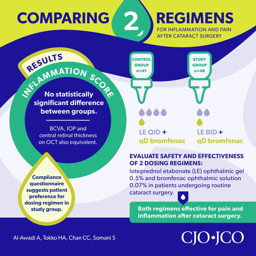
The article is also featured in the CJO’s first Resident Perspectives digest, which highlights a handful of articles from each issue with summaries written by members of the CJO’s Resident Advisory Council. These summaries are written by residents for residents and focus on issues that are particularly relevant to learners here in Canada and beyond.
Post-LASIK : calcul de la puissance des LIO
Mme PKR me dit qu’elle ne s’inquiète pas outre mesure son résultat réfractif après la chirurgie de la cataracte. Mais comme elle a déjà eu une chirurgie réfractive, moi, ça me préoccupe! De toute évidence, c’est une patiente qui se soucie (ou du moins qui s’en est déjà soucié) de la réfraction!
Je pense à un cours sur la cataracte qui se donnait à Toronto en février 2019, où j’ai eu beaucoup de plaisir à rencontrer des collègues, à apprendre de nouvelles idées et à recueillir des perles. Les connaissances que j’ai acquises en cette occasion me seront très utiles pour optimiser les résultats réfractifs chez mes patients en chirurgie de la cataracte.
Nous sommes tous particulièrement conscients des défis liés à la sélection des puissances de lentilles intraoculaires (LIO) chez les patients qui subissent une chirurgie de la cataracte après une chirurgie réfractive. Au fil des ans, nous avons élaboré un certain nombre d’approches différentes pour relever ce défi, mais il reste encore des améliorations à apporter. L’ampleur de ce problème ne fera qu’augmenter à mesure que nous aurons plus de patients qui auront besoin d’une chirurgie de la cataracte après une correction réfractive.
J’aimerais signaler deux liens qui proposent des calculateurs essentiels pour les LIO après intervention réfractive en 2019. Le premier est celui de l’ASCRS et le deuxième, le True K post-LASIK de Graham Barrett. Grâce à ces deux ressources, je crois que bon nombre d’entre nous seront en mesure de nous attaquer plus efficacement à l’avenir à ces cas intéressants.
Calculateur de LIO post-correction réfractive de l’ASCRS : http://iolcalc.ascrs.org/ – N’oubliez pas de choisir le bon type de chirurgie réfractive antérieure, en haut.
Le calculateur Barrett True K : http://www.apacrs.org/barrett_true_K_universal_2/ – Avec celui-ci, assurez-vous de sélectionner le bon type de chirurgie réfractive antérieure dans le menu déroulant.
Recommandé par la Dre Amandeep Rai
Amandeep Rai, MD, FRCSC
Membre du Comité de la carrefour de ressources pour la pratique
Comment améliorer vos connaissances dans la rétinopathie du prématuré (RDP)
Il n’y a pas si longtemps, un résident en ophtalmologie qui était sur le point de terminer son stage en pédiatrie me disait : « Tous les nourrissons que nous avons évalués pour une RDP n’avaient aucune rétinopathie ou avaient une RDP qui ne nécessitait aucun traitement. J’ai peur de ne pas être en mesure de diagnostiquer une RDP de type 1 quand je serai en pratique clinique. Qu’est-ce que je peux faire ? ». La première réponse qui m’est venue à l’idée était de lui recommander de procéder comme je l’ai fait pendant ma résidence : voir plus de patients avec le superviseur de stage et continuer à étudier les critères de la RDP et les photos standardisées. C’est alors que je me suis souvenue d’un super outil de formation par cas sur la RDP qui a été mis en ligne sur le site Web de l’American Academy of Ophthalmology (AAO) lorsque j’étais associée (fellow), et qui s’intitule Retinopathy of prematurity: Case-Based Training (https://www.aao.org/interactive-tool/retinopathy-of-prematurity-case-based-training).
Cet outil interactif en anglais propose 20 cas qui présentent divers degrés de gravité de même qu’un tutoriel, le cas échéant, pour apprendre comment diagnostiquer et quand traiter la RDP. Chaque cas précise le poids de naissance, l’âge gestationnel et l’âge post-menstruel du nourrisson, et fournit six images standard de la RDP pour chaque œil de même que des choix pour établir un diagnostic spécifique et un suivi. Bien que cela puisse sembler tout simple, certains cas vous donneront du fil à retordre, à savoir s’ils répondent véritablement aux critères de mise en route d’un traitement selon l’étude ETROP (Early Treatment of ROP). En fait, certains sont si complexes que les mêmes images et critères ont été soumis aux participants à la conférence récente sur la RDP (http://www.ropupdate.com) – conférence biennale accréditée qui rassemble les plus grands spécialistes en RDP et en néonatalogie et que je recommande vivement) et ont donné lieu à une variété de diagnostics et de schémas de traitement. C’est donc un excellent outil de formation pour les résidents et les associés (fellows), ainsi que pour les spécialistes polyvalents, les pédiatres ou les rétinologues qui font des dépistages de la RDP et qui souhaitent s’assurer que leurs connaissances sont à jour.
Nul besoin d’être titulaire d’un compte de l’AAO pour accéder au module de formation à de seules fins d’apprentissage. Par contre, si vous souhaitez obtenir des crédits d’auto-évaluation pour cette activité, vous devrez être un membre en règle de l’AAO.

Recommandé par la Dre Christine Law
Christine Law, M.D., MSHPEd, FRCSC
Membre du Comité de la carrefour de ressources pour la pratique
Bibliothèque de formation virtuelle en neuro-ophtalmologie
La Neuro-Ophthalmology Virtual Education Library (NOVEL) était l’une de mes ressources pédagogiques préférées en tant que résidente, et demeure une ressource à laquelle je retourne à l’occasion dans le cadre de ma pratique générale. Les cas de neuro-ophtalmologie se dissimulent parfois parmi les cas suspects de vision floue et de glaucome. Chose certaine, ils semblent toujours trouver un moyen de se faufiler dans mes cliniques! Je trouve utile de me rafraîchir la mémoire de temps en temps en neuro-ophtalmologie et, pour moi, une vidéo vaut 1000 mots. NOVEL est un formidable répertoire de documents numériques en libre accès utilisables à des fins d’éducation et de recherche. C’est un excellent endroit où trouver des images et des vidéos pour des présentations, passer en revue les concepts importants en neuro-ophtalmologie et soumettre des cas intéressants.
Le mois dernier, dans le cadre d’un atelier d’introduction à l’ophtalmologie pour un groupe d’étudiants en médecine, j’ai utilisé plusieurs vidéos d’examens cliniques pour présenter les principales techniques d’examen comme l’examen des pupilles et les tests de motilité extraoculaire. Ce site donne aussi accès à toutes les présentations des congrès annuels de la North America Neuro-Ophthalmology Society (NANOS). Dernièrement, je suis tombée sur une belle série de présentations du congrès de 2016, qui passe en revue l’utilisation de la tomographie par cohérence optique en neuro-ophtalmologie.
L’écoute de ces vidéos pourrait facilement devenir un projet d’apprentissage personnel au titre de la section 2 du programme de MDC, qui vous donnerait 2 crédits par heure.
Découvrez NOVEL à l’adresse.
Recommandé par la Dre Anu Mishra
Anu Mishra, M.D., MSHPEd, FRCSC
Membre du Comité de la carrefour de ressources pour la pratique
Non-Infectious Uveitis – What’s Best for my patient: Local or systemic treatment?
The COS is pleased to provide access to the presentations from the co-developed symposium Non-Infectious Uveitis: What’s Best For My Patient: Local or Systemic Treatment?, which took place at the 2018 COS Annual Meeting in Toronto, Ontario. Through these presentations, International and Canadian expert faculty lead a discussion around the various treatment strategies for uveitis, how to choose the most appropriate line of therapy for different uveitis presentations. A novel model of collaborative care between ophthalmology and rheumatology was also presented. Below, you will find a video of the presentation by each speaker.
CPD credits: Scanning resources that are relevant to your professional practice by enhancing your awareness of new evidence, perspectives and findings can be claimed as Section 2: Self-Learning under Scanning in MAINPORT with the MOC Program of the Royal College of Physicians and Surgeons of Canada.
Presentation 1: Use of Anti-VEGFs in Uveitis
Dr. Amin Kherani reviews the epidemiology and pathogenesis of VEGF, then explains the management of uveitis and the role of anti-VEGF therapy.
Presentation 2: Topical and Injected Corticosteroids: When are they appropriate?
Dr. Chloe Gottlieb discusses the appropriate use of topical and periocular steroids for treating uveitis, the steroid-related complications that limit this use, and the benefit of additional therapies.
Presentation 3: Intraocular Implants vs. Immunosuppression
Dr. Marc de Smet covers conventional uveitis medications, and compares immunosuppressive treatment options.
Presentation 4: Immunosuppression for Uveitis: What, when, how, and by whom
Dr. Larissa Derzko-Dzulynsky expands on the area of immunosuppression for uveitis, highlighting first line and second line treatments, including biologics.
Presentation 5: Collaboration Between Ophthalmologists and Rheumatologists for Patients with Uveitis
Dr. Olga Ziouzina presents a collaborative care model for uveitis treatment, drawing from her own experience at a combined multidisciplinary uveitis clinic in Calgary.
Panel Discussion
At the end of the session, moderator and Chair, Dr. Eric Fortin leads the audience and faculty through a series of questions related to Non-Infectious Uveitis: What’s Best for my Patient: Local or Systemic Treatment?
In collaboration with the Canadian Ophthalmological Society, the Canadian Retina Society, and the Canadian Uveitis Society. This symposium was co-developed with AbbVie and was planned to achieve scientific integrity, objectivity and balance.
Cette ressource n’existe qu’en anglais.
CSI: Cataract Surgery Investigation
Symposium Resources
The COS is pleased to provide access to the presentations from the co-developed symposium presented at the 2018 COS Annual Meeting, CSI: Cataract Surgery Investigation. Dr. Ike Ahmed, along with a team of forensic ophthalmologist investigators, presented three different and engaging cases, unveiling clues to analyze the causes of poor outcomes in cataract surgery patients. They discussed tools and strategies to prevent these outcomes from occurring in future patients. Below, you will find video presentations and handouts on complicated cataract surgery.
CPD Credits
Scanning resources that are relevant to your professional practice by enhancing your awareness of new evidence, perspectives and findings can be claimed as Section 2: Self-Learning under Scanning in MAINPORT with the MOC Program of the Royal College of Physicians and Surgeons of Canada.
Episode 1: Residual Sphere
Join forensic expert Dr. Amandeep Rai on his investigation of a spherical refractive error after cataract surgery. Do you have what it takes to uncover the suspect in this refractive surprise?
Episode 2: Astigmatism Post Toric
Special Agent Dr. Rosa Braga-Mele tackles a complicated case of astigmatism post TORIC IOL implant. Can you catch the perpetrator?
Episode 3: The Miniature Ghost
Follow seasoned investigator Dr. Guillermo Rocha as he puts together clues to uncover the truth about an apparent ghost. What will you notice at the dysphotopsia crime scene?
Episode 4: CSI Case Discussions
In the last episode of this season, members of the investigative team field questions and discuss their challenging cases, including the use of different formulas and calculations and the implementation of new technologies.
This symposium was co-developed with the Canadian Ophthalmological Society and Alcon in order to achieve scientific integrity, objectivity and balance.
This resource is only available in English.