


CJO: December 2023 Issue Highlights
The December 2023 CJO is now available online. Here are some of the highlights:
Resident Perspectives + visual abstract: Our talented team of residents have summarized 4 articles with a focus on what’s most relevant to ophthalmology learners here in Canada and around the globe, including the article featured in our October visual abstract, Comparison of pre-formalin fixation and postfixation temporal artery biopsy lengths
Original research articles:
- Updated inventory and projections for Canada’s ophthalmology workforce
- Long-term outcomes of type 1 retinopathy of prematurity following monotherapy with bevacizumab: a Canadian experience
- Assessment of the optic nerve, macular, and retinal vascular effects of COVID-19
- Idiopathic epiretinal membranes: postoperative changes in morphology
- Impact of inherited retinal diseases on Canadian patients and families: a mixed-methods study
- Backlog in ophthalmic surgeries associated with the COVID-19 pandemic in Ontario 2020
Research letters, photo essays, and case reports:
- Antibiotic resistance profile in bacterial keratitis: a single-centre study [research letter]
- Costs associated with treating uveal melanoma are understudied [research letter]
- Hyperviscosity retinopathy from chronic myelogenous leukemia [photo essay]
- Diffuse infiltrating retinoblastoma: a panuveitis masquerade [photo essay]
- Variable phenotypic expression of MYOC mutations in a family with inherited pediatric glaucoma [case report]
- Rapidly progressive optic neuropathy due to optic nerve sheath meningioma following hormonal fertility treatment [case report]
- Optical coherence tomography angular hyperreflectivity as an early sign in acute macular neuroretinopathy [case report]
Follow the CJO on social media:
Facebook: CanJOphth
Instagram: @cjo_jco
LinkedIn: CJO – JCO
Twitter: @CanJOphth
Physician Health & Wellness
Burnout and depression among the medical community is becoming more commonplace. COS has prepared the list below offering our members support on topical resources including webinars, courses, articles, books, and more.
While we all face challenges to varying degrees, our culture of silence in medicine often precludes us from openly sharing what we are facing. Speaking up, however, not only offers you an opportunity to seek support, but also helps others who may be hesitant to speak up for themselves by reminding them they are not alone.
If you are experiencing thoughts and behaviours of depression or suicide, it is important to know that there are immediate supports available to you 24 hours a day. Click here to find a crisis centre near you and seek the support you need.
Resources
Kevin MD (KevinMD.com)
A collection of articles and blog posts on physician suicide and related issues, written by various medical professionals. LEARN MORE
Pamela Wible, MD
A physician and author who advocates for the well-being of medical professionals and patients, with a focus on addressing physician suicide. LEARN MORE
Nina Ahuja, MD FRCSC
Docs in Leadership
An online community for physicians interested in leadership development, emotional intelligence, education, and career development. LEARN MORE
Canadian Medical Association
A New Era in Physician Health and Wellness
A hub for physician wellness resources, including articles, webinars, and tools for promoting well-being and addressing burnout and other challenges. LEARN MORE
Canadian Medical Association
The Silent Stigma Around Physician Suicide
A guide to the signs, risk factors and influences of physician suicide and an overview of the systemic and individual efforts needed to prevent it. LEARN MORE
Canadian Psychological Association
A collection of fact sheets on various mental health topics, including depression, anxiety, and stress, written by psychology experts. LEARN MORE
Canadian Mental Health Association
Workplace Mental Health
A program to promote mental health in the workplace, with resources and tools for employers and employees. LEARN MORE
Not Myself Today
A workplace mental health initiative that provides tools and resources for organizations to promote mental health and reduce stigma. LEARN MORE
Envisioning a Canada Without Suicide
A suicide prevention initiative that aims to raise awareness, provide education and resources, and promote action to prevent suicide and support those affected by it. LEARN MORE
Royal College Peer Coaching in Practice
Peer feedback is an essential element of the Royal College Maintenance of Certification (MOC) Program. While there are many ways to receive peer feedback, one of the most powerful forms of feedback is the type you receive in a peer coaching or mentoring relationship.
This site provides support and resources to guide your personal peer coaching or peer mentoring journey.
CJO: October 2023 Issue Highlights
The October 2023 CJO is now available online. Here are some of the highlights:
Resident Perspectives + visual abstract: Our talented team of residents have summarized 4 articles with a focus on what’s most relevant to ophthalmology learners here in Canada and around the globe, including the article featured in our October visual abstract, OCTA changes following loading phase with intravitreal aflibercept for DME.
Original research articles:
- Investigation of ocular microstructural changes according to disease severity in patients with chronic obstructive pulmonary disease
- Misdiagnosis of fungal infections of the orbit
- Patient perspective on the participation of ophthalmology residents in their cataract surgery
- Cost-effectiveness of locally prepared Descemet membrane endothelial keratoplasty grafts in Edmonton
- Comparison of surgical techniques for recurrent pterygium
- Indications and pathologic diagnoses of diagnostic chorioretinal biopsies in the province of Quebec, Canada
Research letters, photo essays, and case reports:
- Split-detection adaptive optics imaging for cytoid body-like materials in a cotton wool spot [photo essay]
- Polyp clusters: atypical presentation of polypoidal choroidal vasculopathy using OCT angiography [photo essay]
- Intraretinal granuloma in neurocysticercosis [photo essay]
- Accidental intralenticular injection of a dexamethasone implant in an adult male [case report]
- Endoscopy-assisted total pars plana vitrectomy during Boston keratoprosthesis type 1 implantation [case report]
- COVID-19-associated vestibular neuritis in an infant [case report]
- To save the vision of persons with diabetes Canada needs data-informed timely screening [correspondence]
Follow the CJO on social media:
Facebook: CanJOphth
Instagram: @cjo_jco
LinkedIn: CJO – JCO
Twitter: @CanJOphth
The Canadian Ophthalmology Mentorship Program (COMP)

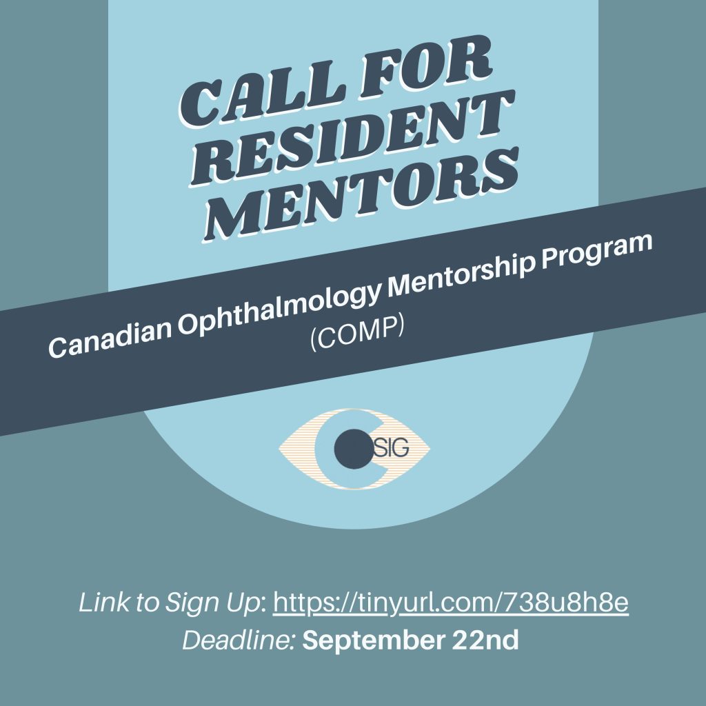
The Canadian Ophthalmology Mentorship Program (COMP) is an annual national mentorship program that connects 3-4 Canadian medical students per ophthalmology resident mentor for the purposes of networking, career exploration, and community building. Recruitment is now open for ophthalmology resident mentors and Canadian medical student mentees.
Sign-ups are open till September 22, 2023.
Link to sign up: tinyurl.com/738u8h8e.
Contact [email protected] with any questions.
Surgical Retina Fellowship program at the Ivey Eye Institute and Western University
Dr. Munir Iqbal is pleased to announce the initiation of a Surgical Retina Fellowship program at the Ivey Eye Institute and Western University in London, Ontario
The fellowship will be 2 years in length, and will include exposure to clinical aspects of retina disease management. The fellow will have the opportunity to evaluate both new and existing patients. The pathology will include but not be limited to: Age-Related Macular Degeneration, Diabetic Retinopathy, Retinal Vascular Diseases, Uveitis, Hereditary Retinal Diseases, Surgical Retinal and Vitreoretinal diseases, including vitrectomy, retinal detachment surgeries, macular surgeries, complex IOL cases, scleral buckling techniques, suprachoroidal drainage, along with pre/post-operative management for surgical cases.
Goals and Objectives:
- To gain exposure and proficiency in the diagnosis and management of medical and surgical retinal conditions to prepare a candidate for a clinical academic career retinal surgery with subspecialty training.
- To obtain training in the appropriate use and interpretation of diagnostic tests, including:
• Fluorescein Angiography
• Optical Coherence Tomography
• Ultrasonography - To develop proficiency in a number of non-surgical procedures, including:
• Laser Photocoagulation
• Photodynamic Therapy (PDT):
• Intravitreal and subTenons injections of pharmaceutical agents: - To develop proficiency in surgical procedures, including:
• Vitrectomy
i. Retinal detachment repair
ii. Macular cases
iii. Vitreous hemorrhage
• Scleral buckling techniques
• Secondary intraocular lens cases - To provide exposure to the evaluation of patients with suspected malignancy, the ancillary testing and evaluation of treatment options and management.
- To develop skills for lecturing to colleagues and residents on various aspects of medical and surgical retinal disease.
- To develop critical appraisal skills and to be aware of current and completed research in the area of medical retinal disease.
Research and Education:
The fellow will be expected to be involved with retina related basic and/or clinical research. Projects will be outlined for the fellow and they will be expected to present this at a national or international meeting. Dedicated time for research will be scheduled into rotation blocks as described above.
The fellow is expected to enhance resident teaching and clinical discourse among the department. Presentation of interesting/educational cases at grand rounds is expected. The fellow will perform 2 grand rounds presentations per academic year.
Application Process:
Interested applicants should send their curriculum vitae as well as 3 recent letters of reference from surgical mentors directly to Dr. Munir Iqbal at [email protected] or Mia O’Neil at mia.o’[email protected]
Applications to the Fellowship Programs must include the following:
• Letter of Intent
• Up-to-date Curriculum Vitae
• 3 Letters of Reference
Eligibility Requirements for Canadian and International Medical Graduates:
• Candidates must possess a medical degree from a University recognized by the Medical Council of Canada (MCC)
• Candidates must have completed an Ophthalmology residency that is recognized by the College of Physicians and Surgeons of Ontario (CPSO)
• International Medical Graduates do not need to write additional exams, but must be approved by the PGME Office at Western University
For More Information and to Register Click here
Cornea Learning Modules
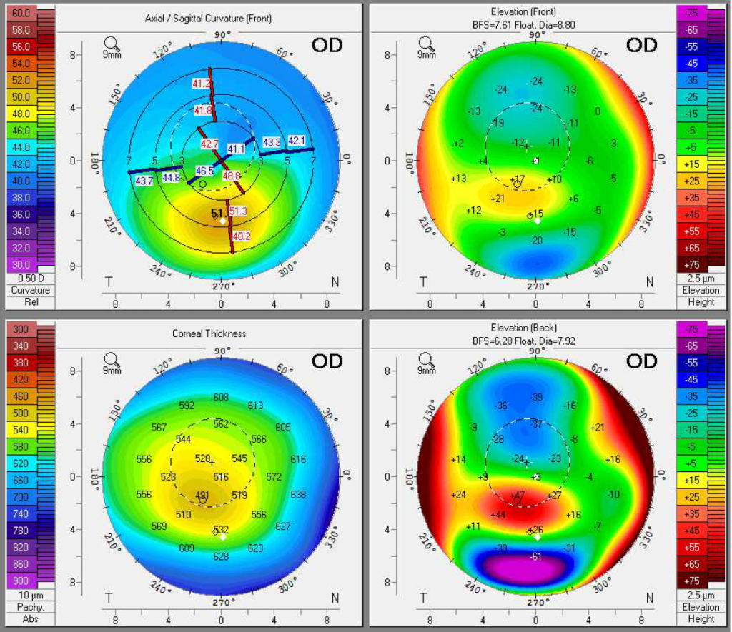
Cornea Learning Modules
This educational program consists of online learning modules of corneal topography and tomography using a case-based format, with the goal of improving knowledge and comfort in interpreting corneal imaging.
Learning Objectives
· Understanding differences between topography and tomography, and which clinical scenarios require use of corneal imaging
· Understand different maps and data presented in topography and tomography, and how they are used in the diagnosis and management of corneal related anomalies
· Interpret corneal imaging in context of normal and diseased eyes (including keratoconus, corneal ectasias, pterygium, assessment prior to cataract or refractive surgery)
CanMEDS Roles
· Medical expert
· Scholar
This is a Member’s Only Section 3 MOC Accredited Activity. You must first log in to your account.
Click here for the Modules Link
This activity is an Accredited Self-Assessment Program (Section 3) as defined by the Maintenance of Certification Program of the Royal College of Physicians and Surgeons of Canada and approved by the Canadian Ophthalmological Society. You may claim a maximum of 2 section 3 credits.
CCOR Fellowship Directory
This is a sortable directory of subspecialty clinical fellowships attended by over 210 Canadian ophthalmology residents. It includes contact information for past residents who indicated they were open to being contacted by prospective fellows.
CCOR Post-residency Guidebook
A Canadian Ophthalmology Resident’s guide to preparing for life after residency. Learn about the career in general vs subspecialty ophthalmology practice, compare different subspecialties, and how to apply for fellowship. Insights shared are a compilation of previous CCOR fellowship talks and tips from a recent survey of graduating residents.