


2025 COS Annual Meeting & Exhibition

Photo credit: Roman Makedonsky
The image used in our annual meeting branding is of a mortuary pole carved by famed Indigenous artist Bill Reid and his assistant Werner True. It can be found in Stanley Park in Vancouver, British Columbia. We encourage our members to visit the pole and learn more about the importance of mortuary poles to the West Coast Haida peoples.
The COS Annual Meeting will take place from June 19 – 22, 2025 in beautiful Vancouver, BC.
Join us at the Vancouver Convention Centre where ophthalmologists and eye care professionals from across the spectrum of vision health, from research to patient care, will come together.
This meeting boasts an outstanding international and Canadian faculty presenting the latest in ophthalmic research and practice. The COS Annual Meeting and Exhibition includes invited lectures, scientific papers, wet labs and workshops, as well as networking opportunities and an extensive exhibition of ophthalmic equipment and services.
The Call for Abstracts is now open, to submit an abstract or find out more, click HERE.
Learning objectives:
By participating in this year’s meeting, attendees will:
- Integrate into their practice, knowledge and skills gained from the sharing of Canadian and international research and scientific studies
- Discuss recent advances in the diagnosis and treatment of eye diseases
- Compare and contrast core concepts, new advances and clinical experiences by networking with colleagues and internationally renowned keynote speakers
- Enhance or develop new skills through innovative hands-on learning experiences in Self-Assessment Program (SAPs) and Surgical Skills Transfer Course (STCs)
- Appraise new and innovative technology and discuss developments in treatment and medical devices with industry representatives in the Exhibition Hall
For more information such as program scheduling, registration, and travel, click HERE.
Cornea Surgical Video Learning Series – Management of Iris Defects: Replace or Repair
Wills Eye Hospital and the University of Toronto are excited to bring you the next installment of the Cornea Surgical Video Learning Series:
Date: Thursday, September 19th 2024
Time: 19:30 – 21:00 EST
Location: Virtual
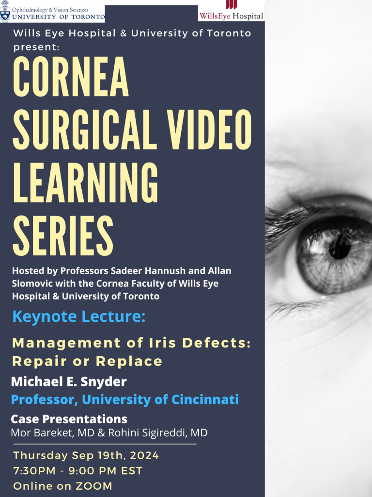
Keynote Presentation:
Professor Michael Snyder, MD, Department of Ophthalmology, University of Cincinnati
Guest panelists: Joshua Teichman, MD FRCSC and Randal Ulate, MD (University of Toronto),
Zeba Syed, MD and Brenton Finklea, MD (Wills Eye Hospital)
Surgery Case Presentations: Rohini Sigireddi, MD (Wills Eye Hospital), Mor Baraket, MD(University of Toronto)
Register in advance for this webinar: https://us02web.zoom.us/webinar/register/WN_ABOjHTL9Rmu7NS9Oh9V1Ag
After registering, you will receive a confirmation email containing information about joining the webinar.
The evening will as usual be recorded and placed online for viewing.
If you would like to visit any of our prior series, please see the link here:
https://ophthalmology.utoronto.ca/subspecialty-rounds
CME credits are available for the following:
· Royal College Maintenance of Certification Section 1: 7.5 hours (1.5 Section 1 hours per session)
· American Medical Association Category 1: 7.5 credits (1.5 Category 1 credits per session)
· European Union for Medical Specialists UEMS-EACCME®: 7.5 credits (1.5 ECMEC credits per session)
CJO: August 2024 Issue Highlights
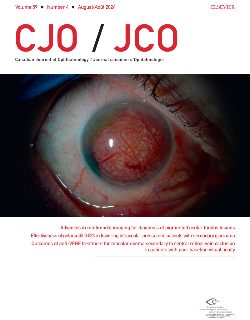
The August 2024 CJO is now available online. Here are some of the highlights:
Resident Perspectives + visual abstract: Our talented team of residents have summarized 3 articles from this issue with a focus on what’s most relevant to ophthalmology learners here in Canada and around the globe. In addition, our August visual abstract provides a visual summary of an article on the Epidemiology of ocular emergencies in a large Canadian eye centre.
Reviews and Original Research Articles:
- Advances in conjunctival melanoma: clinical features, diagnostic modalities, staging, genetic markers, and management
- Advances in multimodal imaging for diagnosis of pigmented ocular fundus lesions
- Research productivity of first-year Canadian ophthalmology residents: a 12-year trend
- Visualization of preretinal membranes using trypan blue in patients with traction retinal detachments
- Artificial intelligence chatbot performance in triage of ophthalmic conditions
- Retinal and choroidal microvascular changes during pregnancy detected with OCTA
Research Letters, Photo Essays and Case Reports:
- Performance of three artificial intelligence chatbots on Ophthalmic Knowledge Assessment Program materials
- Scleral patch graft for emergency open-sky repair
- Cavitary ciliary body melanoma with extensive pigment dispersion
- Acute macular neuroretinopathy following uncomplicated epiretinal membrane removal
- Bilateral circumscribed choroidal hemangiomas in 2 patients: a rare finding
- 532-nm laser for sub-internal limiting membrane hemorrhage associated with retinal macroaneurysms
- Syndromic PRD: case report of McArdle retinopathy and review of literature
Follow the CJO on social media:
Facebook: CanJOphth
Instagram: @cjo_jco
LinkedIn: CJO – JCO
Twitter: @CanJOphth
Interview study on artificial intelligence in ophthalmology
Researchers at LMU Munich are looking for ophthalmology professionals for an online qualitative study on artificial intelligence (AI) in ophthalmology. To understand ophthalmology professionals’ perceptions of AI and to identify relevant factors for a successful integration of AI tools in clinical practice, the research team conducts online interviews (20-25 min) with ophthalmology professionals (e.g., ophthalmologists, ophthalmic nurses/technicians/assistants, optometrists). Participation does not require any previous experience or knowledge of AI. Participants will receive a $20 Amazon gift card and the chance to win another $50 gift card. Ophthalmology professionals interested in participating can access more information and sign up HERE.
Ophthalmology Revision Course
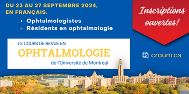
Date: September 23 – 27, 2024
Location: Montreal, QC (in-person)
The Cours de révision en ophtalmologie (CROUM) is a refresher course for French-speaking ophthalmologists and ophthalmology residents from around the world who would like to improve their knowledge. This intensive, 5-day training is being offered by the University of Montreal in Montreal, QC.
The program offers a thorough and modern review of the clinical sciences of ophthalmology, including all of the latest knowledge in the field. While the relevant, basic sciences are covered, the approach remains practical and applicable to your patients. To view more about the program, click HERE.
To view more information about registration for this program, click HERE.
Please note that this course will only be offered in French.
4th Global Neuro-Ophthalmology Case Festival
Date: August 24th – 25th, 2024
Time: 4:00 PM – 9:00 PM IST
The 4th Global Neuro-Ophthalmology Case Festival is a two-day celebration of learning, focused on common, complex, and important neuro-ophthalmology disorders with diverse speakers and expert panelists.
The Case Festival will be available for free on YouTube.
Click HERE to access the August 24th livestream
Click HERE to access the August 25th livestream.
COS 2025 – Call for Abstracts
COS is now accepting abstracts for the 2025 COS Annual Meeting in Vancouver, British Columbia! The abstract submission deadline is Thursday, October 03, 2024 at 11:59 PM ET.
Abstract can be submitted in the following format:
- Paper: oral presentation.
- ePosters: an electronic on-demand ePoster showcased in the Exhibit Hall throughout the Annual Meeting and accessible via the Annual Meeting app.
- Surgical Videos: Show off your surgical skills by submitting a video. Surgical videos will be displayed alongside the ePosters (maximum five (5) minutes in duration).
The COS Annual Meeting historically boasts an 80+% acceptance rate with abstracts making up over 50% of all presentations. With so many abstracts, it is important to set your research apart! Follow these keys to a successful submission when drafting your abstract or editing your video:
- Follow all abstract guidelines; read and understand the rejection criteria.
- List all authors/producers and declare all financial interests for each author/producer.
- Indicate that ethics approval has been obtained, where required.
- Submit your abstract to the appropriate subspecialty category.
- Include a short descriptive title and avoid gratuitous statements and irrelevant information.
- Provide an abstract that is clear, concise, and free of errors.
- Papers and ePosters:
- In the body of your abstract, include study details under the headings: Purpose, study design, methods, results and conclusions. Abstracts with “results pending” or “conclusions to be discussed” will not be accepted. Information about sample size, study duration, follow-up, techniques used, etc. should be included.
- Surgical Videos:
- Submit a video that is well-edited, clear and easy to follow.
- In the body of your written abstract, include an outline of key areas featured in your surgical video.
To learn more and to submit your abstract, follow the link here: https://cos-sco.secure-platform.com/2025/
Understanding Ophthalmologists’ Perspectives on Biosimilars
Background
A needs assessment was conducted to better understand the needs of Canadian ophthalmologists in the context of biosimilars. Biosimilars are biotherapeutic agents that aim to replicate the efficacy and safety profile of their reference biologic counterparts. They undergo rigorous testing to ensure comparable safety, efficacy, and quality. Biosimilars hold the potential to enhance patient access to essential medications and foster competition within the healthcare market.
The purpose of the needs assessment was to determine the necessity of educational programs and other assets to support ophthalmologists in effectively navigating biosimilars.
Ophthalmologists’ Knowledge and Practices
Ophthalmologists are becoming more aware of biosimilars but there is still some hesitancy in adopting their widespread usage. In order to better understand their perspectives, COS conducted 2 surveys – one in English and one in French.
Online surveys were distributed to ophthalmologists across Canada. There was an overall completion rate of 76% (95 complete, 30 partial responses out of 125 total). The English survey had representation from multiple provinces, with the highest being Ontario. The French survey respondents were 100% from Quebec. Despite some provincial skewing, participation was geographically diverse offering a comprehensive view.
The findings revealed that while awareness of biosimilars for anti-VEGF treatments stood at 61.1%, actual usage was notably lower, with only 26.1% of respondents having utilized biosimilars. Preparedness to incorporate biosimilars into practice varied among ophthalmologists, with larger studies, longer-term analyses, and real-world evidence identified as factors that could increase comfort levels with biosimilar adoption.
Interest in educational initiatives was pronounced, with respondents expressing a desire for webinars, online seminars, clinical practice guidelines, and increased educational meetings or journal clubs. However, concerns persisted regarding limited understanding of safety and efficacy, restricted access to information, and uncertainty surrounding regulatory standards and approval processes.
Perceptions and Concerns about Biosimilars
Ophthalmologists voiced nuanced opinions regarding the approval process and the likelihood of biosimilar adoption. Notably, 80% of respondents advocated for implementation of larger clinical trials to ensure the safety and efficacy of biosimilar drugs. However, views on the likelihood of adopting biosimilars in the near future were mixed.
Open-ended responses underscored the diversity of opinions among ophthalmologists, with concerns ranging from safety and efficacy to the potential for cost savings and the necessity for additional evidence-based data. The influence of medication cost and the presence of patient support programs moderately affected decision-making processes. Uncertainty surrounding regulatory standards and approval processes emerged as a common concern.
Conclusions and Next Steps
The needs assessment’s key findings emphasize the imperative for further education, larger clinical trials, and real-world evidence to assuage concerns and enhance ophthalmologists’ comfort levels with biosimilars. These perspectives have tangible implications for decision making processes and the potential adoption of biosimilar drugs in ophthalmology.
Recommendations include the development of comprehensive educational initiatives, collaboration for larger studies, and the generation of real-world evidence to address ophthalmologists’ concerns effectively. Additionally, the establishment of an educational toolkit tailored to biosimilar usage in ophthalmic practice is recommended for guiding informed decision-making and ensuring patient safety.
This needs assessment was completed with an unrestricted educational grant with COS and Apotex, Biocon & Biogen and was planned to achieve scientific integrity, objectivity and balance.
Summary of Data
Awareness and Usage of Biosimilars
- 61.1% of ophthalmologists surveyed are aware of biosimilars for retinal conditions
- However, only 26.1% are currently using them in their practice
Likelihood of Using Biosimilars
- 51% said they are likely to start using biosimilars in their practice in the near future
- 49% said they are not likely to start using them
Factors to Increase Comfort Level with Biosimilars
- More educational meetings/journal clubs (56%)
- Real-world studies (46.2%)
- Longer-term studies (38.5%)
- Larger studies (29.7%)
Educational Interests
Ophthalmologists expressed interest in various educational modalities to increase their knowledge of biosimilars
- Webinars/online seminars (57.1%)
- Clinical practice guidelines (50.5%)
- Peer-reviewed journal and articles (39.6%)
- Case-based discussions/grand rounds (37.4%)
- Mentorship programs (26.4%)
To view the sources for this needs assessment, you can download them here:
We would like to take the opportunity to thank the scientific planning committee for conducting this needs assessment. To view their profiles, you can download the document here:
Video Journal of Cataract, Refractive, & Glaucoma Surgery – The Essential Partnership with Industry (Issue II)

The second issue of the Video Journal of Cataract, Refractive, & Glaucoma Surgery is live! The program is entitled: The Essential Partnership with Industry.
Dick Lindstrom and David Chang give a great introduction detailing the synergy between ophthalmologists and industry that results in innovative products which address unmet needs. For the first time in our 40 year history, we are publishing videos submitted from industry showcasing new products and company philosophies. To view more, please visit: http://www.vjcrgs.com/ and view the table of contents below!