


EyeNovation Webinar: The Light Adjustable Lens – Recording
We are thrilled to announce the next webinar in our “EyeNovation: International Webinar Series for Ophthalmic Expertise” series, “The Light Adjustable Lens: Impressions after 500 and my 2 eyes,” presented by esteemed ophthalmologist Dr. Samir Melki from Harvard Medical School. This webinar took place on October 30th, from 7:00 PM – 8:00 PM ET.
Dr. Melki, an associate professor of ophthalmology at Harvard Medical School, has an impressive background, including completing a fellowship in Cornea and Refractive Surgery at the Massachusetts Eye and Ear Infirmary, an MD PhD from Vanderbilt University, and an Ophthalmology residency at Georgetown University. He is the founder of Boston Vision, an academically-oriented practice, and has published 4 textbooks and 57 articles in respected peer-reviewed scientific journals.
In this webinar, Dr. Melki shared his LAL experience with the first 500 eyes, discussing the technology, process, and outcomes. Attendees will gain insights into:
- The process of the LAL;
- Comparison of the visual outcomes of the LAL and other IOLs;
- Patient satisfaction after LAL implantation.
Dr. Melki also discussed the success rate of monovision after LAL, the percentage of patients requesting a change in refractive aim, and his personal experience with LAL and LAL plus from a surgeon’s perspective.
You can watch the recording below:
Missed this webinar? Stay tuned for future webinars in the EyeNovation series, as we continue to bring you the latest advancements and expert perspectives in ophthalmology. Together, we can improve patient care and push the boundaries of what is possible in this dynamic field.
Vision Health Conference 2024
Report Card: The Post-Pandemic State of Vision Health in Canada
Date: Wednesday, October 30, 2024
Time: 10 am – 2 pm
Location: Toronto Reference Library
789 Yonge Street, Toronto, Ontario, M4W 2G8
Bram & Bluma Appel Salon (2nd floor)
This conference will present results from the newly-released Report Card Part 2, undertaken by the Canadian Council of the Blind (CCB) and Fighting Blindness Canada (FBC), which is a follow-up to the Report Card Part 1, released in October 2022.
The report provides an update on the current state of vision health in Canada and what we can anticipate as we move forward from the post-pandemic era. The report offers insights into the key issues affecting vision health and the community of people living with vision loss.
This is a hybrid event, meaning the same content will be offered at the same time.
For in-person registration, click HERE.
For online registration, click HERE.
You can view the 2023 Report Card below!
27th EVER Congress
Date: November 3-5, 2024
Location: Valencia, Spain – Palacio de Congresos de València
EVER is the leading ophthalmological research association in Europe which covers all areas of ophthalmology and the visual sciences. One of the main activities of EVER is the organizing of a high quality research meeting every year at a location chosen for its access and congress facilities.
The 27th EVER congress will provide the perfect environment to promote the best research in vision, retina and eye, bringing together clinical, translational and basic science in a very beautiful, welcoming and dynamic city.
For more information on this innovative, scientific programming, click HERE.
The 27th EVER CONGRESS, Valencia, Spain 03/11/2024 – 05/11/2024 has been accredited by the European Accreditation Council for Continuing Medical Education (EACCME®) with 17 European CME credits (ECMEC®s). Each medical specialist should claim only those hours of credit that he/she actually spent in the educational activity (6 credits on 3/11, 6 credits on 4/11, 5 credits on 5/11). “Through an agreement between the Union Européenne des Médecins Spécialistes and the American Medical Association, physicians may convert EACCME® credits to an equivalent number of AMA PRA Category 1 CreditsTM. Information on the process to convert EACCME® credit to AMA credit can be found at www.ama-assn.org. Live educational activities, occurring outside of Canada, recognised by the UEMS-EACCME® for ECMEC®s are deemed to be Accredited Group Learning Activities (Section 1) as defined by the Maintenance of Certification Program of the Royal College of Physicians and Surgeons of Canada.
Fuchs IX Symposium
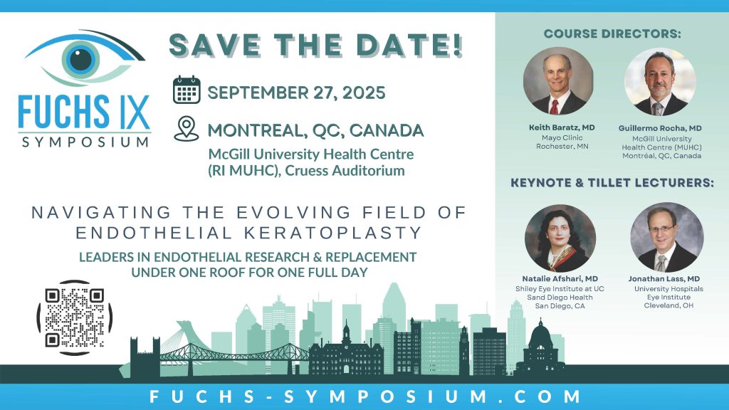
For the past decade, the biennial Fuchs Symposium has brought together the world’s leaders in endothelial research and treatment under one roof to share the latest advancements in this exciting field. The Fuchs IX Symposium will take place at the McGill University Health Centre in Montréal, Canada. The course directors have gathered an incredible faculty that you won’t want to miss. This symposium will take place on September 27th, 2025. Be sure to save the date, registration will be open soon!
CJO: October 2024 Issue Highlights
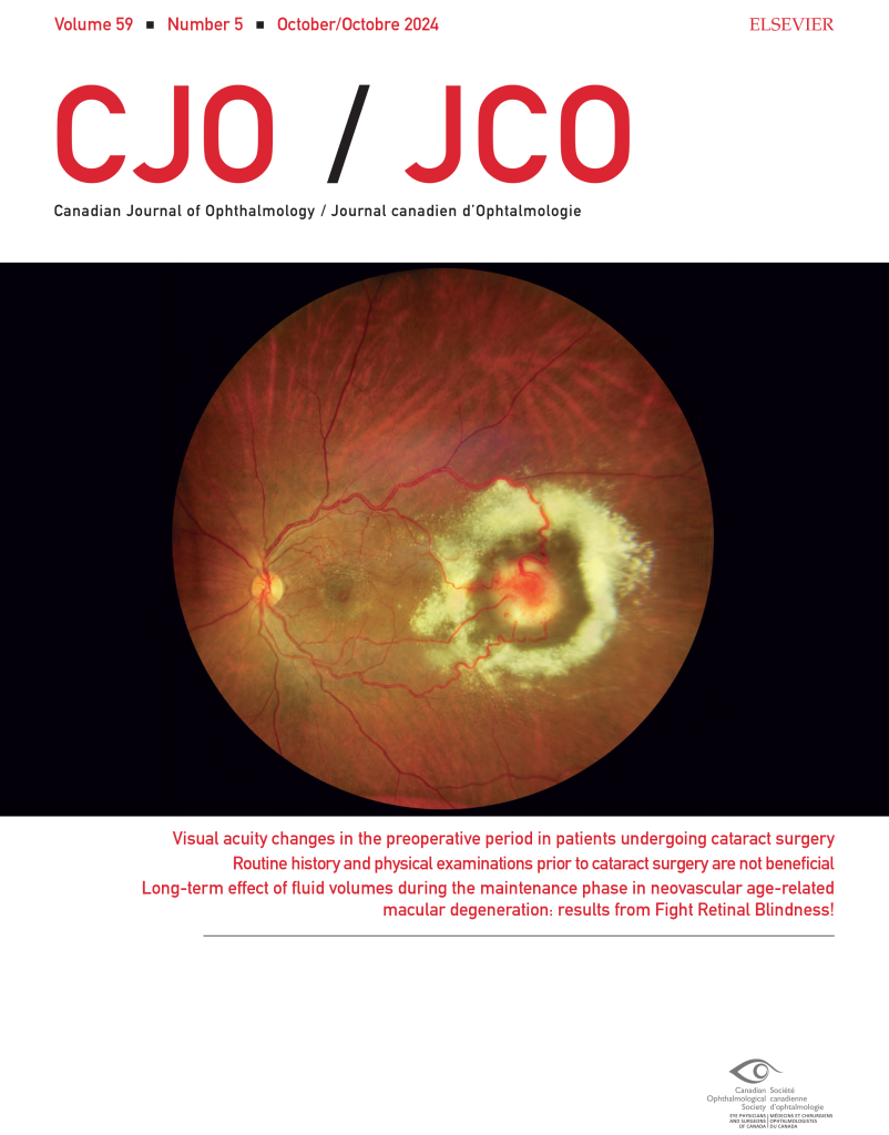
The October 2024 CJO is now available online. Here are some of the highlights:
Resident Perspectives + visual abstract: Our talented team of residents have summarized 3 articles from this issue with a focus on what’s most relevant to ophthalmology learners here in Canada and around the globe. In addition, our October visual abstract provides a visual summary of an article on the Long-term effect of fluid volumes during the maintenance phase in neovascular age-related macular degeneration: results from Fight Retinal Blindness!
Reviews and Original Research Articles:
Research Letters, Photo Essays, Case Reports, and more:
Follow the CJO on social media:
Facebook: CanJOphth
Instagram: @cjo_jco
LinkedIn: CJO – JCO
Twitter: @CanJOphth
World Sight Day – ROP Resource

This year, World Sight Day (October 10, 2024) focuses on children’s eye health around the world. We want to highlight and review the existing guidelines for the screening of retinopathy of prematurity (ROP). These guidelines, developed by key organizations in ophthalmology, aim to ensure early detection and management of ROP in preterm infants to prevent life-long visual impairment and blindness. Please note that this review reflects current clinical recommendations from leading bodies and is not a COS policy or official guideline.
Screening Eligibility
Screening is recommended for infants at the highest risk of developing ROP. The criteria include:
- Gestational age: ≤30 6/7 weeks
- Birth weight: ≤1500 grams
- Neonatal risk factors: Infants born at >1500 grams or >31 weeks may still require screening (eg, infants with hypotension or those who received oxygen supplementation)
Initial screening: The first eye exam is recommended at either 31 weeks’ postmenstrual age in infants with gestational ages less than 26 6/7 weeks at birth, and at four weeks of chronological age in infants born with a gestational ages of 27 weeks or more, whichever is later.
Follow up intervals depend on the severity of the ROP observed in the initial exam.
As we recognize World Sight Day, it is important to remember that infants who develop ROP are at a higher risk of visual disorders such as: strabismus, amblyopia, high refractive errors, cataracts and glaucoma. By raising awareness and ensuring early detection and treatment, we can help protect the vision of these vulnerable children and work towards a world where everyone has access to eye care.
Reviewed by: Dr. Marie-Josée Aubin, MD (Chair), Dr. Cynthia Qian, MD (CPD Council Chair)
References:
Jefferies, A. L. (2016). Screening examination of premature infants for retinopathy of prematurity. https://cps.ca/en/documents/position/retinopathy-of-prematurity-screening
Subramanian, S., Kern, M. D., & Deegan, W. F. III. (2023). Retinopathy of prematurity guidelines. Medscape. https://emedicine.medscape.com/article/976220-guidelines
EyeNovation Webinar: Pearls from the Trenches: What I’ve learned from doing complex cases – Recording
The field of ophthalmology is constantly evolving, with new techniques, technologies, and best practices emerging regularly. To help ophthalmologists stay at the forefront of their profession, we are proud to introduce “EyeNovation: International Webinar Series for Ophthalmic Expertise.”
This innovative webinar series aims to connect ophthalmologists from around the world, fostering knowledge sharing and collaboration. Each webinar will feature renowned experts presenting on cutting-edge topics and sharing their invaluable experiences.
We are thrilled to announce our inaugural webinar, “Pearls from the Trenches: What I’ve learned from doing complex cases,” presented by esteemed ophthalmologist Dr. Steven Safran. With years of experience tackling challenging ophthalmic cases, Dr. Steven Safran will share the lessons he has learned and provide practical insights to enhance attendees’ clinical practice.
The webinar took place on September 30th from 7:00 PM to 8:00 PM EST and included a live Q&A session, allowing attendees to engage directly with Dr. Safran.
By attending this webinar, ophthalmologists will:
- Gain valuable insights from a leading expert in the field
- Learn practical strategies for managing complex ophthalmic cases
- Connect with colleagues from around the globe
- Enhance their clinical knowledge and skills
You can enjoy a recording of the webinar below:
Did you miss this live webinar? Stay tuned for future webinars in the EyeNovation series, as we continue to bring you the latest advancements and expert perspectives in ophthalmology. Together, we can improve patient care and push the boundaries of what is possible in this dynamic field.
Theatre of Medicine
A refreshing new MOC program, delivered by the internationally renowned Shaw Festival, is launching with a 15-hour course this Fall! Come to Niagara-on-the-Lake for this inaugural event, co-created with the Royal College of Physicians and Surgeons of Canada, which focuses on meaningful learning through skills and tools found in the performing arts. The Theatre of Medicine program has been approved for University of Toronto accreditation.
Aligning with emergent quality improvement (QI) guidelines, this unique approach to communication and interpersonal skills training goes beyond patient simulation and role-playing to provide tools that can make an immediate, practical difference.
Learning outcomes will cover:
- Physician Well-being
- Professional Practice
- Interpersonal skills and communication
Looking for more information? Click HERE for details on registration, course information and travel!
55th Sally Letson Symposium
Thank you for attending the Sally Letson Symposium. We greatly appreciate your participation. The 56th Sally Letson Symposium will take place from September 11-13, 2025. Please save the date for next year’s event, and we look forward to welcoming you again!
Join us for the 55th Sally Letson Symposium chaired by Dr. Ian Clark, University of Manitoba, and Dr. Solin Saleh (Co-Chair), University of Ottawa.
This year’s topic is “Pediatric Ophthalmology – Making a difference that lasts a lifetime.” Over the course of 2.5 days, the program will delve into the realm of paediatric ophthalmology. Esteemed guest speakers will deliver captivating lectures and presentations, fostering interactive learning and encouraging critical thinking. Seize the opportunity to enrich your knowledge and skills in this vital field.
Location: Westin Ottawa, 11 Colonel By Drive, Ottawa, ON K1N 9H4
Date: September 19 – 21, 2024
For more information, please visit the Sally Letson Symposium Website.
QUESTIONS? For inquiries relating to the scientific program and CPD credits, please contact Jill Garner [email protected]